
and should have..............
Since Popp clearly showed us all living cells emit ELF-UV.......this phenomena is called fluorescence.
This has huge implications when understanding how sunlight is being changed by the machinery inside of cells to do physiologic work.
Fluorescence is the emission of light by a substance that has absorbed light or other electromagnetic radiation. It is a form of luminescence. In most cases, the emitted light has a longer wavelength, and therefore lower energy, than the original absorbed radiation. This means atomic light emission is highly dependent upon how an atom can absorb light photons.
Absorbing highly powered UVC light would seem to make the most sense for biology since all living cells are known to emit ELF-UV light. Fluorescent bands center at wavelengths longer than the resonance line.
Fluorescence occurs when an atom or molecules relax through vibrational relaxation to its ground state after being electrically excited. The specific frequencies of excitation and emission are highly dependent on the molecule or atom. The energy loss is due to vibrational relaxation while in the excited state. Fluorescent bands center at wavelengths longer than the resonance line. This shift toward longer wavelengths is called a Stokes shift.
Excited states are short-lived with a lifetime at about 10^-8 seconds. Molecular structure and chemical environment affect whether or not a substance luminesces. When luminescence does occur, molecular structure and chemical environment determine the intensity of emission. Generally, molecules that fluoresce are conjugated systems. What is a conjugated state?

The aromatic ring of the aromatic amino acids is an example of a conjugated system. In chemistry, a conjugated system is a system of connected p orbitals with delocalized electrons in a molecule, which in general lowers the overall energy of the molecule and increases stability. It is conventionally represented as having alternating single and multiple bonds.
All aromatic amino acids are photon traps for sunlight.

Why do aromatic side chains all absorb UV light? Why is tryptophan the MOST absorptive of all the aromatic amino acids to UV light?
Remember tryptophan makes melatonin and serotonin and NAD+.
Molecules containing π-electrons or non-bonding electrons (n-electrons) can absorb the energy in the form of ultraviolet or visible light to excite these electrons to higher anti-bonding molecular orbitals. The more easily excited the electrons (i.e. lower energy gap between the HOMO and the LUMO), the longer the wavelength of light it can absorb. There are four possible types of transitions (π-π*, n-π*, σ-σ*, and n-σ*), and they can be ordered as follows: σ-σ* > n-σ* > π-π* > n-π*.”
Now let's look at molecular configuration of tryptophan:
The aromatic rings are composed of pi bonds that absorb UV light. Amino acids like glycine, below, don't have aromatic side chains and therefore do not absorb UV light particularly well.
You might say “yeah, but glycine has a pi bond right there, all amino acids do!” And you're correct, but a single pi bond does not lend itself to UV absorption very well. A conjugated pi system is what gives tryptophan and your skin and eye it's absorptive power.
Tryptophan is critical in quantum thermodynamics of life because melatonin controls mtDNA dynamics of autophagy and apoptosis. Tryptophan is a non-polar aromatic amino acid and it is essential in humans, meaning the body cannot synthesize it. It must be obtained from the diet and it only becomes physiologic when light programs proteins that have tryptophan in it. Tryptophan is also a precursor to the neurotransmitter serotonin, the hormone melatonin, and vitamin B3. Vitamin B3 is niacin. Vitamin B3 is the key fluorophore protein that makes up the electron donor of cytochrome 1 in the NAD+/NADH couple. Tryptophan is also unique in that it is only encoded by the SINGLE codon UGG making it an ideal quasicrystal when hydrated by non acidified water made by mitochondria that can pulse in a variable way with differing powered electromagnetic waves. The frequency of light creates different pulses and it appears these pulses are critical to mitochondrial biology.
Niacin and nicotinamide (B3) are both precursors of the coenzymes nicotinamide adenine dinucleotide (NAD) and nicotinamide adenine dinucleotide phosphate (NADP) in vivo. NAD+ converts to NADP+ by phosphorylation in the presence of the enzyme NAD+ kinase. NADP+ and NAD+ are coenzymes for many dehydrogenases, participating in many hydrogen transfer processes. They do not work with the deuterium isotope of hydrogen either. NAD+ drops in mitochondrial disease states and in normal aging. NAD+ is important in the catabolism of fat, carbohydrate, protein, and alcohol, as well as cell signaling and DNA repair, and NADP+ mostly in anabolism reactions such as fatty acid and cholesterol synthesis. High energy requirements (brain) or high turnover rate (gut, skin) organs are usually the most susceptible to their deficiency. This makes damage to cytochrome one in either the gut, brain, and skin link to many other quantum processes in man. This is very underappreciated in medicine today.
You should remember all UV absorbing molecules all have conjugated ring systems. The ring’s pi electrons absorb the UV light and are stabilized by the surrounding ring. With more places to go, the electrons can be excited without breaking apart the bond. Without the conjugated system, the electrons simply have nowhere to go within the molecule. If they absorb the energy, they leave the orbital and break the bond and no controlled biochemistry could occur.
As you will learn in organic chemistry, molecules that are stable in high energy conformations react quicker and better. It appears this is why life favors them. Stability is largely based on surrounding atoms and their ability to stabilize electrons occupying high energy orbitals.
Conjugated double bond systems are great examples, as are electronegative atoms like fluorine and chlorine when substituted for hydrogen in organic molecules. They pull the electrons towards them and offer stability when an unstable electron arrangement occurs.
Electrons get excited by the light and move up to higher energy orbitals and threaten to destabilize the molecule, but the fluidity of the ring can compensate for that and keep the molecule from breaking apart due to light (photolysis). This allows tryptophan to absorb the energy and hold onto it long enough for your UV-Vis spectrophotometer to register the change in light.
UV and shorter wavelength light (x-rays, gamma rays in the spectrum of light) move electrons in and out of orbitals. UV light of all tyoes only acts on valence electrons in biologic proteins, whereas x-rays and gamma rays have the power to act on core electrons.
A valence electron excited by UV will go to a higher energy orbital. Core electrons are excited by x-rays. Gamma rays can excite core electrons to the point of ejection, and high energy gamma rays can even annihilate the nucleus of an atom.
On the other side of the spectrum, infrared light causes atoms to vibrate as they absorb the energy and this vibration is what causes your food to cook in an oven. Sunburn is a thermal injury to your skin from overdosing on IR-B and C light. In this case, the light energy of the infrared waves is converted to kinetic energy of movement (1535nm) that is related to temperature rise. IR-A is not capable of much thermal injury and this is why it is often called cool heat. IR-A light also happens to be the most dominate ray of light in our sun's light that falls to Earth (42%).
Microwaves cause certain molecules to spin or vibrate, similarly converting light energy into kinetic energy and cooking your food. The water molecule likes to spin when absorbing microwaves and that is what cooks food in a microwave. The glass bowl doesn't have atoms that can spin when exposed to microwaves, so it doesn't heat up along with your food in the bowl.
Remember that electrons ONLY jump energy levels in a discreet manner (quanta), meaning that there must be a certain amount of energy in order for the electron to move to the next orbital. If the energy level of the light is too high or too low the jump won't occur and the light won't be absorbed. Electrons won't just absorb some energy for a while and jump up when the threshold is reached, it's an all or nothing thing. This is how certain wavelengths of light correspond to different electron jumps.
Fluorescence occurs when an atom or molecules relax through vibrational relaxation to its ground state after being electrically excited. The specific frequencies of excitation and emission are dependent on the molecule or atom.
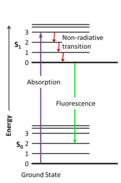
A Jablonski diagram, pictured above and in the video, is used to describe and graph the absorbance, non-radiative decay, and fluorescence of the system. The purple arrow above represents the absorption of light. The green arrow represents vibrational relaxation from singlet excited state, S2 to S1. This process is a non-radiative relaxation in which the excitation energy is dispersed as vibrations or heat to the solvent, and no photon is emitted. The yellow arrow represents fluorescence to the singlet ground state, called So in the picture above.
The fluorescence quantum yield ((\Phi\)) gives the efficiency of the fluorescence process. What defines this? It is the ratio of photons emitted to photons absorbed by the system that defines the quantum yield. Recall all cells release a CERTAIN amount of UV light. This is a big clue what cells are really up to when they are using proteins.
Phi is denoted by the symbol = Φ= # emitted photons divided by the # absorbed photons = quantum yield.
So anyone who lives in sunlight but cannot raise their Vitamin D level normally has a severe form of a quantum yield problem. The same is true if their NAD+ remains low even when they are in the sun even when they eat a ketogenic diet. Food cannot alter a quantum process. This tells us that some other light frequency around them in their environment is affecting their conjugated systems from absorbing UV light. Many illnesses cause this situation and that is why doctors often remark that their patients Vitamin D levels do not appear to budge when inflammatory levels are high. The reason for this in tissues because the absorption coefficient or optical density in their tissues has changed. Water's optical density is known to shift when it is placed in light from Pollack's work. Pollack work is not complete in my opinion.
WHY?
The quantum electrodynamics (QED) description of liquid water structure has been shown by Delguidice that liquid water is a system in a stable non-equilibrium state due to the co-existence of two phases characterized by different thermodynamic parameters: a matrix of non-coherent water molecules hosting many coherence domains. (CDs), about 0.1µm in size, in which all water molecules are oscillating in phase with a self-trapped electromagnetic field. asically, at a fixed temperature and for molecules density exceeding a threshold, the transition of the non-coherent water molecules to the coherence state is spontaneous because it is driving the system to a lower energy configuration - low energy coherent systems (LECS ).
The oscillation of the coherent water molecules takes place between a fundamental state, where electrons are firmly bound (ionization energy of 12.06 mV) and an excited state characterized by a quasi-free electron configuration. The energy of the excited state is 12.06 eV, , which means that only a small amount of energy as (12.60 eV - 12.06 eVV), = 0.54 eV - X is sufficient to extract an electron from biologic systems. X is greater than or equal to 0.1eV and this is the electric potential difference at the CD boundary with the non-coherent water.
This small amount of energy, approximately 0.44 eV, is all that appears necessary for the electron extraction using the oxyhydroelectric effect of EZ water. This makes the coherent water (EZ) act like a reservoir of quasi-free electrons that can be easily released by quantum tunnel effect or by small external perturbation of ambient ELF-UV light from cells to create quantum signaling (quorum sensing). In fact, 0.44 eV is well within the energy that matches the electro-negativity of the O2 molecule.
What does all this imply? Is this is why cells can functon using ELF-UV light to signal all things needed in a cell? Is this how epigenetics really operates? I think it is. This data might solve one of the greatest mysteries in modern biology. Why do cells release this type of light t begin with? Now we know a possible answer. This is how water works in cells with UV light.
Maybe now you can see why all the aromatic amino acids absorb UV light close to 200nm of light which is deep in the UVC range. It appears this is why nature's most critical chemicals have aromatic amino acids in them.
There is a quantum twist here about the absorption spectrum active of aromatic amino acids on Earth.


Fluorescence rarely results from absorption of UV-radiation of wavelengths shorter than 250 nm because this type of radiation is sufficiently energetic to cause deactivation of the excited state by predissociation or dissociation. Most organic molecules have at least some bonds that can be ruptured by energies of this strength. Consequently, fluorescence due to sigma→ σ transitions is rarely observed in nature. Instead, such emission is confined to the less energetic π∗→π π∗→n and the π∗→n transition processes. Fluorescence commonly occurs from a transition from the lowest vibrational level of the first excited electronic state to one of the vibrational levels of the electronic ground state.
Quantum yield (Φ) is greater for π∗→π transition because these excited states show short average lifetimes (larger kf) and because deactivation processes that compete with fluorescence are not as likely to happen in nature. Molar absorptivity of π → π* transitions likelihood is 100-1000 fold greater. The average lifetime is 10^-7 to 10^-9 seconds for n, π* states, respectively.
If every photon absorbed results in a photon emitted. The maximum fluorescence quantum yield is 1.0, and compounds with quantum yields of 0.10 are still considered fluorescent. Another way to define the fluorescence quantum yield is by the excited state decay rates mentioned above and shown in the equations.
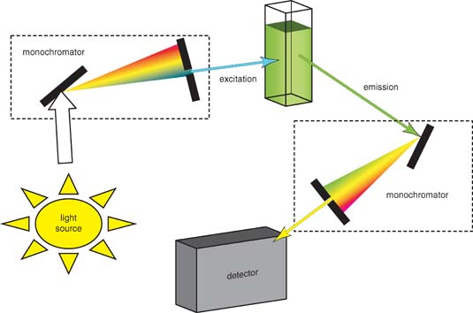
The picture above is a schematic of a typical filter fluorimeter that uses a source beam for fluorescence excitation and a pair of photomultiplier tubes as transducers. The source beam is split near the source into a reference beam and a sample beam. The reference beam is attenuated by the aperture disk so that its intensity is roughly the same as the fluorescence intensity. Both beams pass through the primary filter, with the reference beam being reflected the reference photomultiplier tube. The sample beam is focused on the sample by a pair of lenses and causes fluorescence emission. The emitted radiation passes through a second filter and then is focused on the sample photomultiplier tube. The electrical outputs from the two transducers are then processed by an analog to digital converter to compute the ratio of the sample to reference intensities, which can then be used for qualitative and quantitative analysis. To obtain an emission spectrum, the excitation monochromator is fixed and the emission monochromator varies. To obtain an excitation spectrum, the excitation monochromator varies while the emission monochromator is fixed.
Fluorescence spectroscopy can be used to measure the concentration of a compound because the fluorescence intensity is linearly proportional to the concentration of the fluorescent molecule. Fluorescent molecules can also be used as tags. For example, fluorescence in situ hybridization (FISH) is a method of determining what genes are present in an organism's genome. Single-stranded DNA encoding a gene of interest is covalently bonded to a fluorescent molecule and washed over the organism's chromosome, binding to its complementary sequence.
The presence and placement of the gene in the organism then fluoresces when shined with ultraviolet light. Green fluorescence protein (GFP) is used in molecular biology to monitor the activity of proteins. The gene encoding GFP can be inserted next to a gene encoding a protein that will be studied. When the genes are expressed, the protein will be attached to GFP and can be identified in the cell by its fluorescence.
If cells emit ELF-UV light it means that cells must have built a novel way of creating a spectrum of UV light stronger than UVB and UVA light. That process was covered in the Vermont 2018 video. If you cannot create that light, based on all the fancy science above it shows you that fluorescences in your proteins will be ALTERED and physiology will change. This is especially true with respect to melatonin and NAD+ levels. Technology causes this effect in humans. This is why they cause diseases and increasing our aging with a chronic technology abuse.
Dr. Jack Kruse
2023-02-27 20:11:04 +0000 UTCDr. Jack Kruse
2019-03-02 14:33:33 +0000 UTCJason Gookin
2019-03-01 18:03:47 +0000 UTCDr. Jack Kruse
2019-02-27 22:52:57 +0000 UTCMyster Spock
2019-02-27 18:20:22 +0000 UTCjulia pace
2019-02-21 15:07:20 +0000 UTCDr. Jack Kruse
2019-02-20 01:50:24 +0000 UTCDr. Jack Kruse
2019-02-19 21:04:03 +0000 UTCDiego Fant
2019-02-18 14:21:57 +0000 UTCMonique itzhakov
2019-02-18 08:42:17 +0000 UTCDr. Jack Kruse
2019-02-18 04:27:42 +0000 UTC